What Causes Moya Moya disease?
The cause of moya moya disease is not well understood. It is more common in Japan than the United States, but it is unclear is this is due to environment or genetics. Moya moya disease has been seen in association with a variety of genetic conditions such as: tuberous sclerosis, sickle cell anemia, neurofibromatosis, fibromuscular dysplasia and Down’s syndrome. It is also seen in children who have received radiation to treat some types of brain tumors.
How Is Moya Moya Disease Diagnosed?
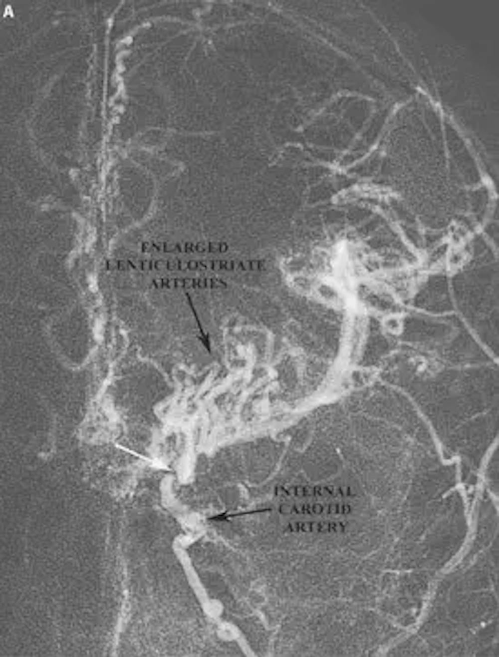
Moya moya disease is typically encountered in two groups of patients: 1) Children less than ten years of age and 2) Adults in their third decade of life. Moya moya disease in children typically presents with non-hemorrhagic strokes and seizures. In the adult population, however, moya moya more commonly presents with brain hemorrhages. Cerebral aneurysms are also frequently seen in patients with moya moya disease, and the rupture of the aneurysms may account for the brain hemorrhages in some patients. Once suspected, the diagnosis of moya moya disease is confirmed with a cerebral angiogram. The angiogram often shows occlusion of the carotid arteries with a small network of blood vessels at the base of the brain resembling a “puff of smoke.”
How Is Moya Moya Disease Treated?
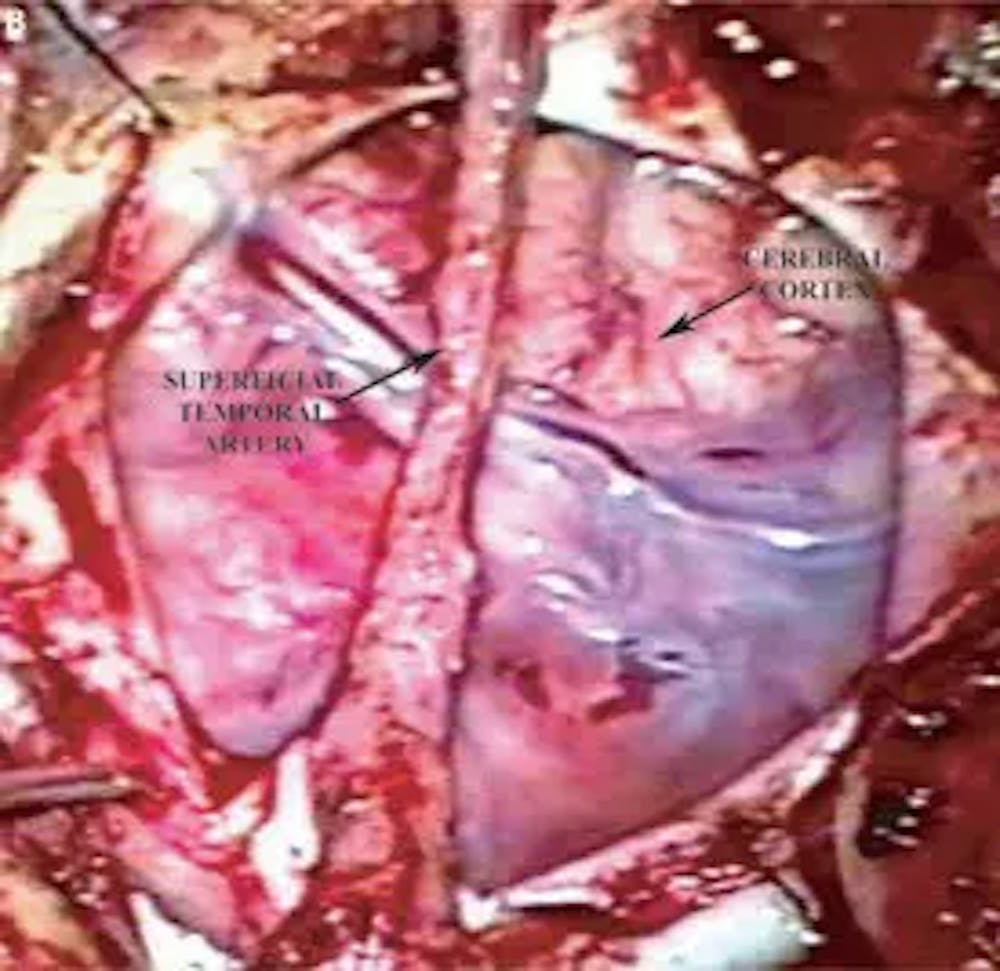
Since the primary problem with moya moya disease is impaired blood flow to the brain, the treatment centers around restoring cerebral blood flow. One method of restoring blood flow is to perform an intracranial-extracranial bypass procedure. Other methods of improving blood flow, such as encephalomyosynagiosis (EMS) or encephaloduroarteriosynangiosis (EDAS), involve placing a layer of muscle or scalp on the surface of the brain, helping to stimulate the growth of new blood vessels in the brain over time.

A) Pre-operative AP angiogram demonstrating the characteristic findings of moya-moya disease. Note the ragged appearance of the internal carotid artery as well as the region where the internal carotid artery is occluded (white arrow). Also note the very large lenticulostriate arteries that resemble wisps or puffs of smoke rising into the air.

B) Intra-operative photo of an encephaloduroarteriosynangiosis (EDAS). In this procedure a vascularized pedicle of tissue supplied by the superficial temporal artery is placed on the surface of the brain. Over time this vascularized tissue will simulate the formation of new blood vessels in the brain.
