A type II chiari malformation is a more complex anomaly usually associated with spina bifida. A type II chiari malformation involves both abnormalities of the bone, cerebellum, and brain stem at the cervico-medullary junction. With a type II chiari malformation, the brainstem has an unusual shape, and a large portion of the cerebellum descends through the foramen magnum. A type III chiari malformation is an uncommon and more extreme variant associated severe neurological deficits. Other conditions sometimes associated with chiari malformations include hydrocephalus, syrinx formation, and spinal curvature (scoliosis).
What Causes a Chiari Malformation?
Although some type I chiari malformations can be acquired following drainage of spinal fluid from the lumbar spine, this is uncommon. The vast majority of all chiari malformations are attributed to structural problems that arise during development.
How Are Chiari Malformations Discovered?
A type I chiari malformation can be associated with a spectrum of symptoms. Type I chiari malformations can be associated with severe headaches that classically are aggravated by coughing. Some patients with type I chiari malformations may note weakness in the arms and legs as well as a variety of sensory disturbances. Some patients will even note difficulty walking, talking or swallowing.
A type II chiari malformation may be associated with severe difficulty breathing and swallowing. Some patients with type II chiari malformations may experience arm weakness and even quadraparesis.
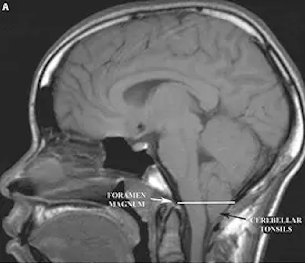
If a chiari malformation is suspected, the diagnosis is confirmed with an MRI of the brain and spinal cord. An MRI is the best way to demonstrate the anatomic anomalies associated with these malformations.
How Are Chiari Malformations Treated?
Type I chiari I malformations with mild or no symptoms may be observed. However, surgery is the only method to treat the symptoms associated with both type I and II chiari malformations. The surgery aims to relieve the pressure placed on the brainstem and spinal cord by the low lying cerebellar tissue. Almost all patients with a type II chiari malformation have hydrocephalus (the accumulation of cerebrospinal fluid in the brain itself) requiring the placement of a shunt.

A) Sagittal T1 weighted MRI of the brain demonstrating the protrusion of the cerebellar tonsils below the foramen magnum (indicated by the white line). This is a type I Chiari malformation.

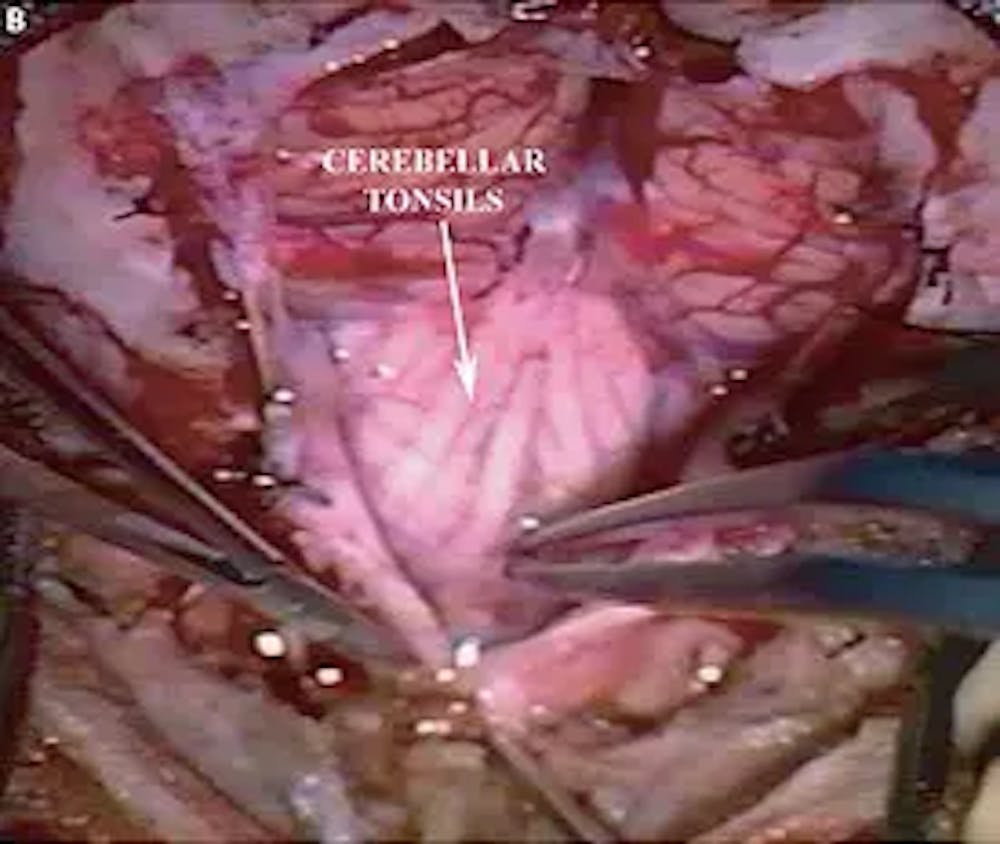
B) Intra-operative photo demonstrating the cerebellar tonsils.

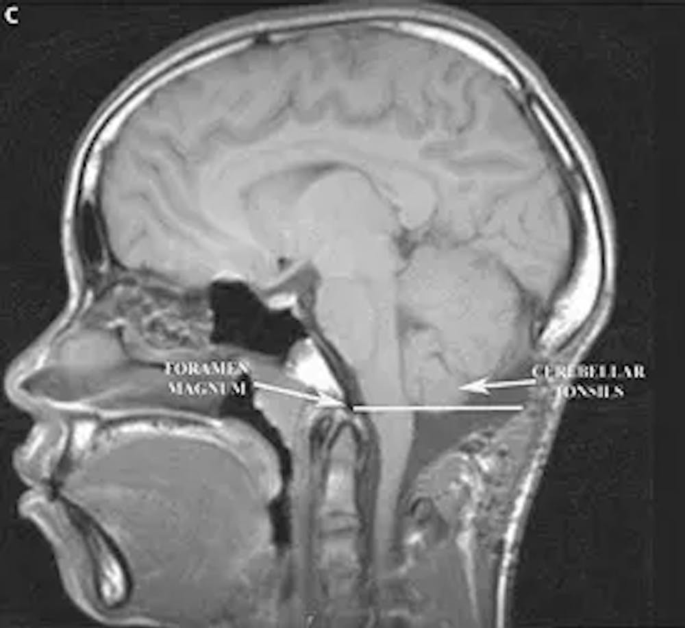
C) Post-operative sagittal T1 weighted MRI of the brain demonstrating the new position of the cerebellar tonsils above the foramen magnum (indicated by the white line)

