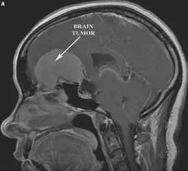
- Meningiomas arise from the arachnoid coverings of the brain.
- Astrocytomas arise from the fibrous support sells in the brain called astrocytes.
- Oligodendrogliomas arise from cells called oligodendrocytes which produce myelin for the nerves of the central nervous system.
- Ependymomas arise from the ependymal cells that line the fluid containing spaces of the brain (ventricles).
- Schwannomas arise from schwann cells that produce myelin covering the cranial nerves.
- Dermoids and epidermoids arise from abnormally located developmental remnants of skin tissue located within the brain.
- Pituitary tumors arise from cells that make up the pituitary gland.
Most primary brain tumors can be further classified into four grades established by the World Health Organization (WHO). Grade I tumors are benign and surgically curable while Grade IV tumors exhibit very aggressive behavior. A pilocytic astrocytoma is a type of astrocytoma that is benign and surgically curable (WHO grade I). A glioblastoma multiforme (GBM) is an type of astrocytoma that is highly invasive with a rapid rate of growth (WHO grade IV).
What Causes a Brain Tumor?
Many brain tumors have genetic abnormalities that alter their patterns of growth. Some genetic disorders are associated with an increased risk of brain tumor formation, such as neurofibromatosis, Von Hippel-Lindau disease, or retinoblastoma. Exposure to extremely high doses of radiation increases the risk of brain tumor formation.
How is a Brain Tumor Diagnosed?
Most commonly a patient will complain of headache occasionally associated with blurry vision and nausea or vomiting. Some patients may present with new onset seizure activity. Brain tumors, however, may produce a variety of symptoms depending on their location in the brain, for example:
- A tumor located in the region of the brain that controls arm or leg movement will result in weakness.
- A tumor in the region of the brain associated with speech will result in language and word finding problems.
- A tumor in the region of the brain that controls balance the patient may complain of unsteadiness and dizziness.
- A tumor in the frontal lobes of the brain may result in personality changes.
Once a brain tumor is suspected, imaging of the brain such as an MRI or CT scan with contrast is critical for confirming the diagnosis and for formulating further treatment strategies.
How Is A Brain Tumor Treated?
Some combination of biopsy, surgical resection, radiation therapy and chemotherapy is employed in the treatment of brain tumors. The exact treatment strategy depends on the exact type of brain tumor. Before any treatment plan can be established the type of tumor should be confirmed by either biopsy or surgical resection.
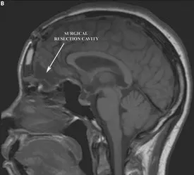
Surgical resection is the mainstay in the treatment of most brain tumors. Surgical resection accomplishes several goals: it establishes a tissue diagnosis for the tumor, it relieves pressure that the tumor is placing on surrounding brain, and it decreases the number of tumor cells in the brain potentially making additional radiation and chemotherapy more effective. Biopsy is usually reserved for obtaining a tissue diagnosis when the tumor is located in a surgically inaccessible part of the brain. Depending upon the tumor type additional chemotherapy and radiation may be required.
A) Pre-operative sagittal T1 MRI with contrast demonstrating a large meningioma compressing the frontal lobes
B) Post-operative sagittal T1 MRI demonstrating resection cavity and resolution of mass effect on the frontal lobes


